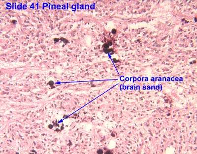
رمل الدماغ[1] (acervuli)[2][3] أو الجسم الرملي[4] (corpus arenacea)[5] هي تراكيب مُتكلسة في الغدة الصنوبرية ومناطق أخرى من الدماغ مثل الضفيرة المشيموية. تمتلك الكائنات الحية القديمة العديد من الجينات العميقة، وإن وجدت فإنَّ وظيفتها غير معروفة. تزداد تركيزات رمال الدماغ مع تقدم العمر، حيث تصبح الغدة الصنوبرية مرئية بشكل متزايد في الأشعة السينية مع مرور الوقت، وعادة في العقد الثالث أو الرابع. تستخدم أحيانًا كمعلمٍ تشريحي أثناء الفحص الإشعاعي.[6]
أظهر التحليل الكيميائي أنها تتكون من فوسفات الكالسيوم (تتميز فيما بعد بهيدروكسيل أباتيت[7]) وكربونات الكالسيوم وفوسفات المغنسيوم وفوسفات الأمونيوم.[8] في الآونة الأخيرة، وصفت رواسب الكالسيت كذلك.[9]
المراجع
- "Al-Qamoos القاموس - English Arabic dictionary / قاموس إنجليزي عربي". www.alqamoos.org. مؤرشف من الأصل في 03 يناير 20203 يناير 2020.
- Vígh, B; Szél, A; Debreceni, K; Fejér, Z; Manzano e Silva, MJ; Vígh-Teichmann, I (1998). "Comparative histology of pineal calcification". Histology and Histopathology. 13 (3): 851–70. PMID 9690142. مؤرشف من الأصل في 17 أكتوبر 201604 أغسطس 2016.
- Kim, Jinkyung; Kim, Hyun-Wook; Chang, Soeun; Kim, Jee Woong; Je, Jung Ho; Rhyu, Im Joo (2012). "Growth patterns for acervuli in human pineal gland". Scientific Reports. 2: 984. Bibcode:2012NatSR...2E.984K. doi:10.1038/srep00984. PMC . PMID 23248747.
- "Al-Qamoos القاموس - English Arabic dictionary / قاموس إنجليزي عربي". www.alqamoos.org. مؤرشف من الأصل في 3 يناير 20203 يناير 2020.
- Tomonari, Yuki; Sato, Junko; Wako, Yumi; Tsuchitani, Minoru (2012). "Age-related Histological Findings in the Pineal Gland of Crl:CD(SD) Rats". Journal of Toxicologic Pathology. 25 (4): 287–91. doi:10.1293/tox.25.287. PMC . PMID 23345933.
- "thesis:The Effect of Fluoride on the Physiology of the Pineal Gland" ( كتاب إلكتروني PDF ). مؤرشف من الأصل ( كتاب إلكتروني PDF ) في 15 ديسمبر 201904 أغسطس 2016.
The calcified pineal is used as a landmark in skull X- rays because of its radio-opacity
- Angervall, Lennart; Berger, Sven; Röckert, Hans (2009). "A Microradiographic and X-Ray Crystallographic Study of Calcium in the Pineal Body and in Intracranial Tumours". Acta Pathologica et Microbiologica Scandinavica. 44 (2): 113–119. doi:10.1111/j.1699-0463.1958.tb01060.x.
- Bocchi, Giancarlo; Valdre, Giovanni; Valdre, Giovanni (1993). "Physical, chemical, and mineralogical characterization of carbonate-hydroxyapatite concretions of the human pineal gland". Journal of Inorganic Biochemistry. 49 (3): 209–20. doi:10.1016/0162-0134(93)80006-U. PMID 8381851.
- Baconnier, Simon; Lang, Sidney B.; Polomska, Maria; Hilczer, Bozena; Berkovic, Garry; Meshulam, Guilia (2002). "Calcite microcrystals in the pineal gland of the human brain: First physical and chemical studies". Bioelectromagnetics. 23 (7): 488–95. doi:10.1002/bem.10053. PMID 12224052.
مراجع إضافية
- Garma-Aviña, A. (2000). "Excretory Plugs from the Choroid Plexus in the Cerebrospinal Fluid of Dogs with Neurological Disease: Possible Role in the Formation of Corpora Arenacea". Journal of Comparative Pathology. 123 (2–3): 146–51. doi:10.1053/jcpa.2000.0405. PMID 11032668.
- Reiter, Russel J.; Welsh, Marcia G.; Vaughan, Mary K. (1976). "Age-related changes in the intact and sympathetically denervated gerbil pineal gland". American Journal of Anatomy. 146 (4): 427–31. doi:10.1002/aja.1001460405. PMID 941859.
- Klein, Pavel; Rubinstein, Lucien J (1989). "Benign symptomatic glial cysts of the pineal gland: a report of seven cases and review of the literature". Journal of Neurology, Neurosurgery & Psychiatry. 52 (8): 991–5. doi:10.1136/jnnp.52.8.991. PMC . PMID 2677249.
- Vigh, B; Vigh-Teichmann, I; Aros, B (1989). "Pineal corpora arenacea produced by arachnoid cells in the bat Myotis blythi oxygnathus". Zeitschrift für Mikroskopisch-anatomische Forschung. 103 (1): 36–45. PMID 2756745.
روابط خارجية
- صور نسيجية: 14401loa — نظام تعلم علم الأنسجة في جامعة بوسطن
- صورة تشريحية: 41_03 في مركز جامعة أوكلاهوما للعلوم الصحية. - "الغدة الصنوبرية"